


GIDORA is the next-generation CT reconstruction software application developed by iLAND based on its many years of know-how and experience. Including a technique for removing metal artifacts, the biggest problem facing Dental CT diagnoses, GIDORA offers a variety of features such as precise CT value output, various correction techniques and many more that dramatically improve the reliability of the CT system. Diagnoses using RevoluX + GIDORA will make a striking change to everyday practice.
1Removing Metal Artifacts
The largest barrier to CT diagnoses is metal artifacts. iLAND’s efforts all started with a simple question “Can we remove metal artifacts fundamentally, not making them unseen by image processing?” After years of trial and error, iLAND has finally established a unique technique for removing metal artifacts to solve this long-standing problem.
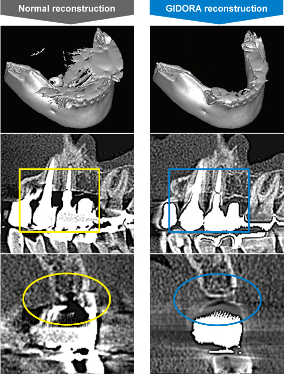
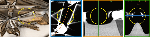
During CT imaging, a metal shape can be recognized on raw data (projected data) in pre-reconstruction state (pfoto below). Focusing on this phenomenon, iLAND has developed an algorithm that automatically recognizes metal shapes in the raw data. This dramatically eliminates metal artifacts and makes it possible to diagnose tooth quality, jawbones and teeth roots that were impossible to see before.
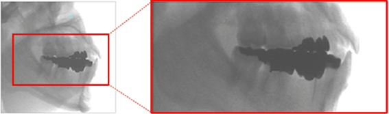
By looking at the raw data shown on a detector, a metal shape can be recognized, such as an inlay or a crown or the like.
GIDORA does not use an interpolation technique.
To say that diagnoses are now possible where it was not possible to see before often gives the impression that the image of invisible area is generated virtually by calculations using linear interpolation from the vicinity image data. However, GIDORA does not use such interpolation techniques.
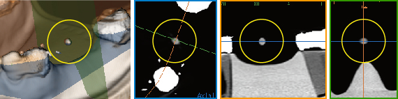
When CT-imaging ceramic sphere (Photo above) that floats between crowns, in a normal reconstruction, it would be completely invisible because of the effects of the metal artifacts generated by crowns (upper of photo below). When the same data is reconstructed using GIDORA, no use of interpolation techniques make it possible to examine the previously invisible ceramic spheres (lower of photo below).
|
Normal reconstruction Ceramic sphere between crowns cannot be seen at all. |
 |
|---|---|
|
GIDORA reconstruction Ceramic sphere is visible. |
 |
2Precise CT Value Output
Why CT values for dental CTs are not outputted like those for medical CTs? It has been. iLAND has solved this long-standing despair with precise CT value output. Clinical bone quality diagnoses are now possible with dental CTs.
Medical CTs were invented in the latter half of the 1960's by Dr. Hounsfield and at the same timing, the concept of CT values (Hounsfield units) was also born. This concept is considered to be the definition of a CT device in today's practice. With air at -1000, and water at 0, density values are given to each individual pixel in response to gray scale. This makes quantitative diagnoses possible regarding the kind of tissue and whether any changes have occurred in the tissue. Particularly in dental practices, not only being able to see the shape and structure of bones in the CT image, but clinical bone quality diagnoses are also possible where softness and hardness of the bones is visualized by coloration according to the CT values.
iLAND considers precise CT value output as No.1 priority. Through repeated research and development efforts, iLAND has achieved highly accurate CT value outputs even for dental CTs.
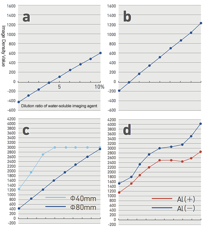
a and b are linear, but the water value is not shown as 0.
For c, not only water is not shown as 0, but the density values vary according to the capturing modes. Image density values peak part way in a narrow imaging range mode.
d does not indicate that water is 0, and has no linearity. Furthermore, numerical values dramatically vary depending on whether there is an aluminum rod in the imagine visual field. This device can be considered not to have density absoluteness. Also, the expression range on the Y axis for c, and d is twice that of a medical CT.
-
CT value of GIDORA, Medical CT and monochrome X-ray
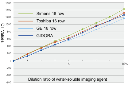
Comparision of CT value for three types of medical CT and GIDORA
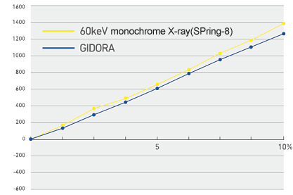
Comparision of CT value for monochrome X-ray* and GIDORA
*Measured at 3rd generation Super photon ring facility(SPring-8)
-
Coloration based on CT value
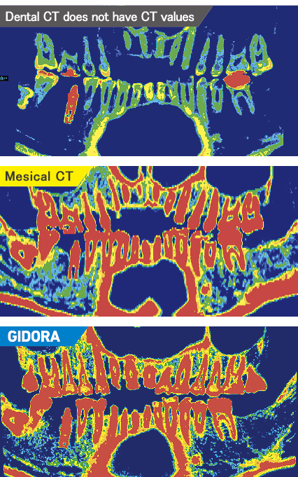
Highly accurate CT values enable clinical bone quality diagnosis
With GIDORA, highly accurate CT values can be outputted that are equivalent to medical CTs and CT values measured by the third generation Super Photon Ring facility (also known as SPring-8).
Also, by using coloring based on the CT values, clinical bone quality diagnoses are possible that are equivalent to medical CTs.
Obtaining high-accuracy CT Values
Does the dental CT output accurate image density, even though it has a different structure to that of the medical CT? iLAND has implemented various correction techniques to be able to brush this concern away.
The reliability of dental CTs has been improved to the level of medical CTs by implementing iLAND’s various technical corrections such as ‘Beam Hardening Correction’ which is indispensable to attain high-accuracy image density. In addition to that, ‘Protrusion correction’ and ‘Scattered Radiation Correction’ solve problems that are unique to dental CTs, and the like.
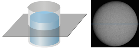
CT imaging of container of water. Cut at horizontal plane to measure CT value of water at diameter portion(Blue line) on same CT image.
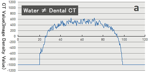
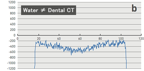
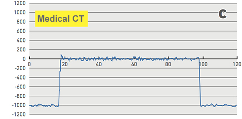
When looking at a profile traversing XY planes of a phantom, although there is some noise, water is shown to be substantially horizontal near 0 on the medical CT, [(c)]. However, the water profiles of the dental CT are not shown as 0 [(a) and (b)]. Not only that, there is greater noise than on a medical CT and a convex portion is formed at the top on most devices [(a)]. In rare cases, there are devices that shown a convex portion below. [(b)]
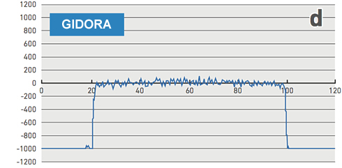
When configured using GIDORA, there is only little noise component, and water is shown to be substantially horizontal near 0. At the air portion on the edge, -1,000 is shown, indicating that the proper corrections were implemented in the same way as with a medical CT. [(d)]